Brief Introduction to the Eye
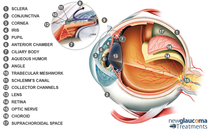
For an organ that is about the size of a large gumball, the eye is a complicated structure. Fortunately, a basic understanding of glaucoma and it’s treatments can be gained with an awareness of just a few “parts” of the eye.
How the Eye Works
It’s helpful to think of the eye as a video camera hooked up to a TV. A video camera has a lens to focus an image, film or a sensor to capture or record the image, and a cable to transmit the image to a TV.
Your eye works in a similar way. It has two surfaces that focus an image: the cornea and the natural lens of the eye. The cornea, which is a clear, curved surface, bends light passing through the eye. The lens fine-tunes the light before passing it onto the retina, located at the back of the eye. The retina records the light, much like film in a camera, and then converts it into an electrical signal. From here, the signal is transmitted to your brain through the optic nerve, which is similar to the cable that connects a video camera to a TV. The brain then processes the signal to let you see the image.
Now let’s take a look at each of the basic parts of the eye and what they do.
Parts of The Eye
Sclera
The sclera is the “white of the eye”. It functions as the outer protective layer of the eye.
Conjunctiva
The conjunctiva is the clear membrane that covers the sclera. It looks a bit like plastic wrap with small blood vessels in it. Because it is clear you don’t generally notice this membrane unless you have an irritated eye, allergies, an eye infection, or have eye surgery. Any of those conditions will cause the blood vessels to become dilated. This results in the conjunctiva changing its appearance from a thin, transparent tissue to a red, thickened blanket over the sclera.
Cornea
The cornea is the clear dome-like structure at the front of the eye. Contact lenses are placed on the cornea. LASIK is a surgery that reshapes the cornea to improve vision. As you might guess from that, the role of the cornea is to focus light as it enters the eye. One of the things that helps to focus light is the tear film, a thin layer of fluid that covers the front surface of the cornea. If the tear film is disrupted then vision can be affected.
Iris
Behind the cornea sits a disc-shaped spongy muscle called the iris. When people discuss eye color they are really talking about the color of the iris. The most common iris colors are brown, hazel, green, and blue. Some glaucoma medications can change the color of the iris.
Pupil
In the center of the iris is an opening called the pupil. Light enters the back of the eye through the pupil which appears like a black circle in the middle of the iris. When we are in the dark, the pupil enlarges to allow more light into the eye. This also happens when we are scared, excited, or aroused.
Anterior Chamber
The anterior chamber is simply the fluid-filled space between the back side of the cornea and the front of the iris. Many glaucoma surgeries are performed in this tiny space.
Ciliary Body
The ciliary body is located just behind the iris. It is made up of little finger-like projections that produce a fluid called the aqueous humor.
Aqueous Humor
The front part of your eye is filled with a clear fluid (intraocular fluid or aqueous humor) produced by the ciliary body. This fluid provides essential nutrients and other chemicals required by the various tissues in the eye. After leaving the ciliary body, this fluid flows around the pupil and into the eye’s drainage system through what is called the “angle.” The fluid then exits the eye through a grate-like structure called the trabecular meshwork where it is then absorbed into the bloodstream through the eye’s drainage system.
Angle
Where the cornea meets the iris a natural angle is formed. The apex (tip) of this angle is where most of the “drainage system” of the eye is located.
Trabecular Meshwork
At some point the aqueous fluid must leave the eye. It does so though a grate-like structure called the trabecular meshwork. At least in normal eyes the trabecular meshwork appears to be the main point of resistance to fluid exiting the eye.[1] Thus, most glaucoma treatments have focused on addressing this part of the eye.
Schlemm’s Canal
This is a small tubular structure that sits beside the trabecular meshwork and encircles the eye. It functions as the natural drain of the eye.
Collector Channels
From Schlemm’s Canal the aqueous fluid moves into small channels. These channels direct this fluid into the bloodstream.
Lens
Like the cornea, the function of the lens is to focus light. The lens is approximately the size and shape of an M&M®. Unlike an M&M®, however, the lens is generally clear (unless it has become a cataract). The lens is suspended behind the iris by microscopic cables called zonules. Light enters the center of the lens through the pupil. When light exits the lens it should be focused onto the retina.
Retina
The retina is the “screen” onto which light is focused. If the cornea and lens are clear and of the correct power then this image should be in focus. Unlike a movie screen, however, the retina does not display an image. Rather, it captures the light and converts it into signals the brain can interpret giving us the ability to see the world around us.
Optic Nerve
All of the focusing and capturing of light would be of no use if this information could not find its way to the brain. The function of the optic nerve is to do just that – transmit a signal from the eye to the brain. This cable-like structure is composed of over a million fibers that pass from the retina to various parts of the brain.
The part of the optic nerve which can be seen by your ophthalmologist in the exam room is called the optic nerve head. It is approximately 1.8mm in diameter on average. Damage to the optic nerve can often be appreciated by your ophthalmologist and described by various terms such as “cupping” or “pallor”. Cupping is a central depression in the optic nerve head commonly seen in those with glaucoma. It indicates that nerve fiber has been lost.
Choroid
This is a soft, vascular (filled with blood vessels) tissue that sits between the retina and the sclera. The choroid may fill with fluid or blood after certain types of glaucoma surgeries.
Suprachoroidal Space
This is a “potential” space between the choroid and the sclera. Usually the choroid and the sclera are sandwiched immediately next to each other. This space may have a role in allowing aqueous to exit the eye.
References
- Grant WM. Further studies on facility of flow through the trabecular meshwork. AMA Arch Ophthalmol. 1958;60(4 Part 1):523-33.
Grant WM. Experimental aqueous perfusion in enucleated human eyes. Arch Ophthalmol. 1963;69:783-801. - Lujan BJ, Wang F, Gregori G, et al. Calibration of fundus images using spectral domain optical coherence tomography. Ophthalmic Surg Lasers Imaging 2008;39(suppl): S15–20.
Don’t delay getting checked for glaucoma.
Make an appointment with an eye doctor in your area now. If you live in the greater Los Angeles area and would like Dr. Richardson to evaluate your eyes for glaucoma call 626-289-7856 now. No referral required. Appointments are available, Tuesday through Saturday.


