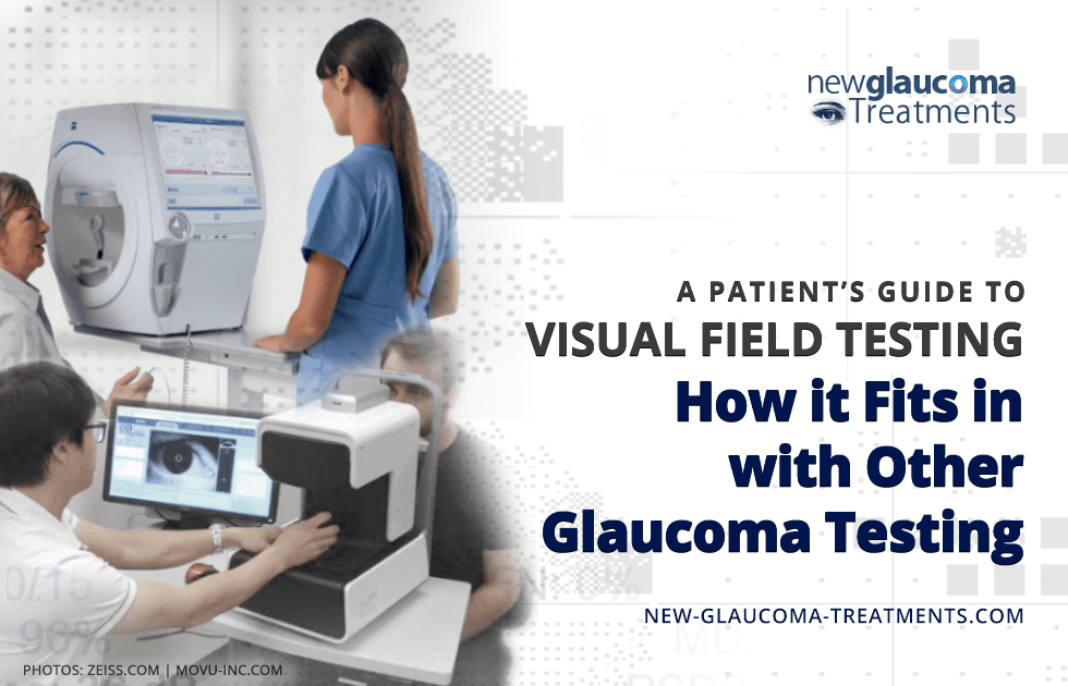
A Patient’s Guide to Visual Field Testing

Reasons Why Visual Field Testing Alone is Inadequate to Follow Glaucoma Progression
As important as visual field testing may be, it is not the only test that should be performed in most individuals being followed for glaucoma. There are numerous reasons for this but the main ones are based on sensitivity and reliability.
Sensitivity
In medicine we use the term “sensitivity” to convey how likely it is that a given test will find what we’re looking for. A test that has 100% sensitivity will find a condition (assuming it is there) every time. Unfortunately, automated perimetry is not very sensitive. Evidence suggests that as much as a third of the nerve tissue damaged by glaucoma can be lost before visual field loss is detected[1]. Thus, at least for early detection of glaucoma, visual field analysis is insufficient for both discovery and monitoring of disease progress.
Reliability
As medical testing goes, automated perimetry is one of the least reliable. If it were a car, it’d be a Yugo (remember those?). Nonetheless, if you’re only other option to get from “A” to “B” were a horse and buggy, you’d take the Yugo. Automated visual field testing has to be performed for the reasons I’ve stated elsewhere. But it suffers from being a subjective test (dependent upon the responses of the individual being tested) that can be negatively impacted by one’s environment, attention, and alertness, among other criteria.
It’s clear that we cannot rely solely on visual field testing to detect and monitor glaucoma. Fortunately we do have other methods that are able to shore up some of the weaknesses of threshold visual field testing.
Optic Nerve Head Examination
Almost as despised as visual field testing is the dilated eye examination. It’s long, mildly uncomfortable, and leaves one with blurred vision for hours after the exam. Nonetheless, it is a key component of monitoring for glaucoma. A skilled physician can detect early signs of glaucoma long before it becomes evident on visual field testing. Additionally, there are signs such as bleeding on the surface of the optic nerve that may portend worsening of glaucoma[2] and that cannot be detected by any means other than direct examination or photography. Even photography tends to be of better quality when the eye is dilated.
Optic Nerve Head Scanning
One of the most exciting developments in the detection and monitoring of glaucoma is (OCT). Briefly an OCT scanner measures the optic nerve head shape and the thickness of the retinal nerve fiber layer (RNFL) that runs from the retinal surface through the optic nerve to the brain. As glaucoma worsens it thins out the RNFL. OCT testing is accurate to about 4 micrometers (four millionths of a meter)! As such, it can detect very small changes in the RNFL that would suggest the presence of glaucoma.
How Optic Nerve Head Scanning
Compliments Visual Field Testing
My own patients often (and reasonably) ask me, “Why do I need both visual field and optic nerve head scanning?” After all, testing can be expensive, time consuming, and frustrating. There should be good justification for whatever testing is recommended by your physician. In the case of glaucoma, I will often use the analogy of how a mechanic figures out what may be wrong with a car. My father was a mechanic and I think I may have learned as much about diagnosing from him as I did from my medical school professors. When searching for what is wrong with a car both a physical examination of the engine is necessary as well as a test drive. Both provide information the other cannot. A physical inspection tells you what might be worn down or disconnected whereas the test drive tells you how the car is underperforming. In the case of OCT scans and visual field testing, the OCT scan is the inspection and the automated perimetry is the test drive. Neither one alone is sufficient to diagnose or monitor glaucoma.
When Visual Field Testing is Inferior to
Optic Nerve Head Scanning
Optic nerve head scanning has the potential to detect glaucomatous damage before visual field loss could be detected[3]. As such OCT scanning of the RNFL may be superior to visual field testing in those with suspected or early glaucoma.
When Visual Field Testing is Superior to
Optic Nerve Head Scanning
As glaucoma progresses, RNFL loss often correlates with loss of visual field, but this is not always the case[4]. Additionally, measuring the RNFL is only useful for detecting thinning or change in early to moderate glaucoma. In those with advanced glaucoma there is little value in scanning the optic nerve head. Thus, visual field testing takes on greater importance as glaucoma severity increases.
When Visual Field Testing may not be Necessary
If glaucoma is a concern and the individual can perform a reliable visual field test, then periodic testing should be performed. However, there are times when it may be reasonable to follow glaucoma without visual field testing. Examples include any condition that sufficiently limits one’s ability to obtain a quality visual field test, such as the following:
- Inability to sit upright for at least 5 minutes
- Neck (cervical) disorders that make it difficult or painful to properly position the head against the chin rest and head rest
- Severe arthritis of the hands
- Severe dry eye
- Dementia or other cognitive disorder that limits attention
- Vision that is too poor to see even the largest and brightest of test stimuli
Summary
Visual field testing, though a mainstay of glaucoma detection and monitoring, has a number of weaknesses which can be addressed through obtaining complimentary testing such as OCT. Indeed, combining visual field testing and OCT measurements of the retinal nerve fiber layer is rapidly becoming the standard of care in modern glaucoma practices.
(This will take you to Dr. David Richardson’s Website)
References:
[1] Kerrigan-Baumrind LA, Quigley HA, Pease ME, et al. Number of ganglion cells in glaucoma eyes compared with threshold visual field tests in the same persons. Invest Ophthalmol Vis Sci.
2000;41(3):741–748
[2] Airaksinen PJ, Heijl A. Visual field and retinal nerve fibre layer in early glaucoma after optic disc haemorrhage. Acta Ophthalmol. 1983;61(2):186–194
[3] Bowd C, Zangwill LM, Berry CC, et al. Detecting early glaucoma by assessment of retinal nerve fiber layer thickness and visual function. Invest Ophthalmol Vis Sci. 2001;42(9):1993–2003.
[4] Armaly MF. The correlation between appearance of the optic cup and visual function.
Trans Am Acad Ophthalmol Otolaryngol. 1969;73(5):8

David Richardson, MD
Medical Director, San Marino Eye
David Richardson, M.D. is recognized as one of the top cataract and glaucoma surgeons in the US and is among an elite group of glaucoma surgeons in the country performing the highly specialized canaloplasty procedure. Morever, Dr. Richardson is one of only a few surgeons in the greater Los Angeles area that performs MicroPulse P3™ "Cyclophotocoagulation" (MP3) glaucoma laser surgery. Dr. Richardson graduated Magna Cum Laude from the University of Southern California and earned his Medical Degree from Harvard Medical School. He completed his ophthalmology residency at the LAC+USC Medical Center/ Doheny Eye Institute. Dr. Richardson is also an Ambassador of Glaucoma Research Foundation.


