A Patient’s Guide to Visual Field Testing

Quick Review of the Current Limitations of Visual Field Testing
Today, automated threshold testing is most commonly used to detect loss of vision from glaucoma. It is based on technology that is decades old. Although software improvements have been made to speed up the testing and better detect changes in the visual field, it is essentially the same testing that was done a half century ago.
Some of the issues of currently available automated perimeters that need to be addressed include:
- Testing Location
- Positioning
- Testing requires a response from the person being tested
- The testing process is boring
Part 1: Virtual Reality (VR) Visual Field Testing
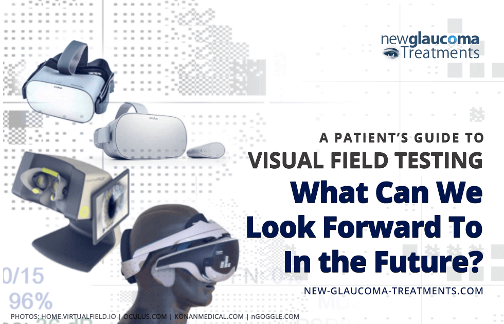
Virtual Reality (also known as “VR”) headsets are capable of displaying images on our visual field. They are relatively light and can be comfortably worn on the heads of most every adult. Early headsets as well as the more powerful modern headsets tend to be tethered by a cable to a powerful computer. Newer headsets, however (such as the Oculus Go) are self-contained and portable so can be used anywhere. This combination of display ability and wearability has the potential to address a number of the issues that currently limit modern automated perimetry.
VIDEO: Dr. David Richardson trying out the now available Virtual Field visual field testing device. Notice how he is able to sit back in the chair in comfort while he is being tested. Contrast that to someone seated upright and leaning forward during standard visual fields testing.
Testing Location
One of the major limitations of currently available visual field testing is the need for the individual with glaucoma to come into the doctor’s office. Even when ones personal schedule does not conflict with the doctor’s office schedule, this is both inconvenient and time consuming. In-office automated perimeters are large and not at all portable so taking one home is out of the question. Virtual reality headsets such as the Oculus Go, however, are very portable. One could foresee a time when such headsets could be loaned out to patients with glaucoma for daily or weekly at-home visual field testing. Alternatively, for those who already own VR headsets a visual field program could be downloaded to their own headset and the results could be sent to their eye doctor.
Positioning
One of the limitations of currently available automated perimeters is the need to position the patient up against a chin and forehead rest. Those who are particularly short or tall or have back or neck disease may not be able to sit comfortably against this rest for the time it takes to complete the testing. Virtual reality headsets, however, can be placed on anyone who can maintain an upright head position. This would potentially allow anyone who can sit upright (even those who are bedridden) to complete visual field testing.
Testing requires a tactile response
Current visual field testing devices require that a handheld controller be pressed whenever a light is seen in the field of vision. There are at least two problems with this:
- Not everyone has good hand-eye coordination and there is a limited period of time after the light object appears during which one has to press the button for it to be counted as “seen” by the perimeter. As we get older what is termed our “reaction time” gets longer. This is made even worse by conditions that slow our mental activity such as dementia.
- It is difficult for some to depress the button on the controller. For example, those with arthritis experience this issue.
Virtual Reality headsets have the ability to track our head movements. This makes it possible to eliminate a handheld controller. Instead of pressing a button, the individual is instructed to look toward the stimulus by moving the head. This is both natural and easy for almost everyone to do with the exception of those with cervical (neck) disease.
The testing process is boring
As I mentioned elsewhere, automated perimetry is essentially like playing the world’s most boring video game. Even compared to “pong” (a variation of the very first video game), visual field testing just doesn’t compete. It’s thought that if automated perimetry weren’t so boring, the quality of the testing would be improved. As such, there is a movement to “gamify” visual field testing. Essentially, the test would be more interactive and enjoyable than what is done today. Early work has been done with virtual reality headsets with encouraging results. However, this work will have to be validated against current threshold testing to see if it can really be used to follow those with glaucoma.
References:
[1] Kerrigan-Baumrind LA, Quigley HA, Pease ME, et al. Number of ganglion cells in glaucoma eyes compared with threshold visual field tests in the same persons. Invest Ophthalmol Vis Sci.
2000;41(3):741–748
[2] Airaksinen PJ, Heijl A. Visual field and retinal nerve fibre layer in early glaucoma after optic disc haemorrhage. Acta Ophthalmol. 1983;61(2):186–194
[3] Bowd C, Zangwill LM, Berry CC, et al. Detecting early glaucoma by assessment of retinal nerve fiber layer thickness and visual function. Invest Ophthalmol Vis Sci. 2001;42(9):1993–2003.
[4] Armaly MF. The correlation between appearance of the optic cup and visual function.
Trans Am Acad Ophthalmol Otolaryngol. 1969;73(5):8

David Richardson, MD
Medical Director, San Marino Eye
David Richardson, M.D. is recognized as one of the top cataract and glaucoma surgeons in the US and is among an elite group of glaucoma surgeons in the country performing the highly specialized canaloplasty procedure. Morever, Dr. Richardson is one of only a few surgeons in the greater Los Angeles area that performs MicroPulse P3™ "Cyclophotocoagulation" (MP3) glaucoma laser surgery. Dr. Richardson graduated Magna Cum Laude from the University of Southern California and earned his Medical Degree from Harvard Medical School. He completed his ophthalmology residency at the LAC+USC Medical Center/ Doheny Eye Institute. Dr. Richardson is also an Ambassador of Glaucoma Research Foundation.


